Osteosarcoma is a bone cancer primarily affecting children and young adults, typically in long bones like the femur, tibia, and humerus. As the most common malignant bone tumor, it is aggressive and can metastasize, making early detection crucial. The cancer originates from osteoblasts, disrupting normal bone structure, and its rapid spread requires prompt intervention. While it makes up about 3% of childhood cancers, its aggressive nature necessitates comprehensive management. Genetics and environmental factors are thought to contribute, guiding preventative strategies and improving early detection.
The Role Of CT Scans In Osteosarcoma Detection
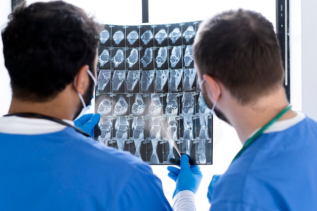
Computed Tomography (CT) scans are crucial in detecting and assessing osteosarcoma, offering detailed cross-sectional images that allow for a thorough evaluation of bones and surrounding tissues. CT scans are invaluable in determining the size, location, and spread of the tumor, helping to distinguish between benign and malignant bone lesions. This differentiation is vital for treatment planning and predicting outcomes. Additionally, CT scans play a key role in surgical planning by providing precise tumor localization, aiding surgeons in removing cancerous tissue while preserving healthy bone. This makes CT scans essential in the comprehensive management of osteosarcoma.
Benefits of Early Detection with CT Scans
Early detection of osteosarcoma significantly enhances treatment outcomes, and oncology imaging, particularly through CT scans, plays a crucial role in this process. CT scans provide highly detailed cross-sectional images, enabling clinicians to identify bone tumors at an earlier stage when they are more localized and manageable. According to oncology imaging specialists, this precision allows for prompt intervention, which can lead to more favorable prognoses.
Detecting osteosarcoma early often means that patients can undergo less invasive treatments. Tumors caught before metastasis tend to respond better to chemotherapy and radiation, reducing treatment intensity and side effects. Additionally, early-stage tumors can often be surgically removed with greater accuracy, preserving surrounding healthy tissue and maintaining limb function.
CT scans, as part of a broader oncologic imaging toolkit, are therefore instrumental not just in detecting cancer but in shaping personalized and effective treatment strategies that improve both survival rates and quality of life.
How CT Scans Work: A Detailed Explanation
CT scans use X-ray technology to create detailed cross-sectional images of the body. During the procedure, the patient lies on a table that slides into a doughnut-shaped machine, capturing images from multiple angles. These images are processed into slices or a 3D representation of the area being examined. CT scans are particularly useful for differentiating between tissue types like bone, muscle, and fat, offering a comprehensive view of osteosarcoma and its impact on surrounding structures. However, the benefits must be weighed against the risks, such as radiation exposure.
Signs And Symptoms Of Osteosarcoma
Recognizing osteosarcoma symptoms is key for early diagnosis. Common signs include persistent bone pain, swelling, and tenderness that worsen at night or with activity. A lump or mass on the bone may develop, sometimes mistaken for a sports injury or growing pains, especially in children. Other symptoms include unexplained weight loss, fatigue, and fever, which require medical attention. Early detection, combined with imaging like CT scans, aids timely diagnosis and improves outcomes.
CT Scan Procedures: What To Expect
A CT scan is generally a straightforward and comfortable process when patients are informed. Before the scan, patients may need to change into a gown and remove metal objects. During the procedure, they lie on a motorized table that slides into the scanner and must remain still for clear images. The process is painless, lasts 10 to 30 minutes, and may involve contrast dye to improve tissue visibility. Patients should inform their provider of any allergies or medical conditions to ensure a smooth experience.
Limitations Of CT Scans In Osteosarcoma Diagnosis
While CT scans are crucial for osteosarcoma detection, they have limitations. One concern is exposure to ionizing radiation, which increases the risk of secondary cancers, especially in younger patients. Additionally, CT scans may struggle to differentiate between soft tissues, requiring other imaging modalities like MRI for better contrast. Although CT scans can assess the tumor’s size and extent, they don’t provide detailed biological information, making biopsy and histopathological analysis necessary for confirming the diagnosis. Despite these limitations, CT scans remain a vital tool in osteosarcoma diagnosis.
Treatment Options Following Early Detection
Treatment for osteosarcoma depends on factors like tumor size, location, stage, and the patient’s health. A multidisciplinary approach is used to optimize outcomes. Surgery is central, with limb-sparing procedures preferred to preserve mobility. If not feasible, amputation may be necessary for complete tumor removal. Chemotherapy and radiation therapy are commonly used alongside surgery. Chemotherapy targets cancer cells, while radiation uses high-energy rays to destroy them. These treatments may be given before (neoadjuvant) to shrink the tumor or after (adjuvant) to eliminate remaining cancer cells.
Integrating CT Scans Into Routine Screening For Osteosarcoma
Integrating CT scans into routine screening for osteosarcoma could improve early detection and outcomes, particularly for high-risk groups such as those with a family history or genetic predispositions. Early identification of asymptomatic tumors allows for timely intervention, reducing the need for aggressive treatments and enhancing survival rates and quality of life. However, the risks of radiation exposure must be considered. Effective screening programs require collaboration among healthcare providers, researchers, and policymakers, along with educational efforts to raise awareness of osteosarcoma and the importance of early detection. By including CT scans in routine screening, we can enhance detection and improve patient outcomes.
Conclusion: The Importance Of Early Detection And Treatment
In conclusion, CT scans are essential for the early detection and management of osteosarcoma, providing detailed images that enhance diagnosis and treatment planning. Early detection through CT scans can lead to less invasive treatments and improved surgical outcomes. Despite limitations like radiation exposure and challenges in differentiating soft tissues, CT scans remain a crucial diagnostic tool. When included in routine screening, they facilitate early identification, enabling timely interventions and better outcomes. As we advance diagnostic technologies, promoting early detection and the use of CT scans will help reduce the impact of osteosarcoma and improve the lives of those affected.